Intraventricular Flow Mapping
We have developed echocardiographic imaging algorithms – iVFM (intraventricular vector flow mapping) – to build intracardiac velocity vector fields from color Doppler. The original iVFM algorithm [IEEE Trans Med Imaging, 2010] is integrated in some Fujifilm Healthcare and Esaote ultrasound machines. We have then improved our algorithm based on a constrained regularization approach, with automated parameter selection [Phys Med Biol, 2021]. The latest version of iVFM is guided by neural networks at each step: endocardium segmentation, dealiasing, vector reconstruction [IEEE Trans Ultrason Ferroelectr Freq Control, 2024].
|
Click to enlarge |
Click to enlarge |


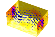
Intraventricular flow mapping becomes three-dimensional using triplane Doppler echocardiography [Phys Med Biol, 2022].

Click to enlarge
Ultrafast Echocardiography
We have developed duplex echocardiography (i.e., B-mode + tissue Doppler) at over 400 frames/s using diverging waves. To achieve this, motion compensation was integrated into coherent summation of signals, to account for phase delays generated by myocardial motions [IEEE Trans Med Imaging, 2016]. From this ultrafast echocardiography technique, we have also developed a least squares method, combining tissue Doppler and optical flow, to obtain myocardial velocity maps with high temporal resolution [IEEE Trans Med Imaging, 2018].

Click to enlarge
Biomechanical Modeling
Biomechanical models, and/or in vitro experiments, are developed for technical and/or clinical studies, to develop and validate new ultrasound imaging techniques or new diagnostic parameters. For example, we analyzed and validated, in patients, the effect of aortic stenosis on coronary flow reserve (CFR) using a lumped parameter model [J Appl Physiol, 2009]. We also develop advanced modeling such as SPH ("smoothed particle hydrodynamics") coupled with a linear acoustic model to simulate color Doppler and vector Doppler images [Phys Med Biol, 2018].

Click to enlarge
Clinical Studies
Based on hemodynamic principles or new imaging approaches, new clinical parameters are proposed to provide better diagnosis in patients with heart disease (e.g., aortic stenosis, diastolic dysfunction, coronary stenosis). In particular, we proposed the energy loss index to improve the assessment of aortic stenosis [Circulation, 2013]. We also prospectively verify whether the CFR/FFR ratio can better guide coronary intervention [PLoS ONE, 2019].

Click to enlarge
Miscellaneous

- RF signals are usually sampled at 4 times the central frequency
- Undersampling of RF signals can be effectively used in ultrasound imaging
- This can avoid data overload and facilitate transfer
- See article #53

- Diverging wave ultrasound requires coherent combination of signals
- Neglecting myocardial motion causes destructive interference
- Compensating for motion preserves coherence and yields quality images
- See articles #41, 50, 52, 54
- SMOOTHN is an automatic, fast, and robust smoother based on DCT
- This automatic smoother can handle outliers and missing values
- The Matlab code was selected as "MATLAB Central Pick of the Week"
- See articles #24, 27, 29

- A large blood vortex forms in the left ventricle during diastole
- Its kinematic and dynamic properties reflect diastolic function
- This vortex can be clinically deciphered by color Doppler echocardiography
- See articles #26, 33, 45, 46

- To simulate color Doppler, we used SPH
- Fluid particles also act as ultrasound scatterers
- The coupled simulator is 3D compatible and easily parallelizable
- See article #55

- Cardiac speckle tracking evaluates regional myocardial function
- Our goal: provide accurate velocities at high frame rates
- We developed a method based on optical flow and tissue Doppler
- See article #52